Atlas
Human skin. 25 year old man
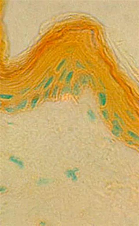
X 500. Acid phosphatase reaction, 60 minutes. (© 1995 Vevy Europe Photoimage)
Cutaneous crest. The acid phosphatase activity is localized in the same places as in the case of fig.1 and with the same intensity. In green tint, you can see the nuc leous of thee cells stained with methyl green.
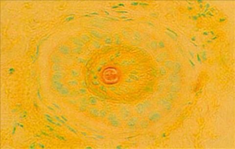
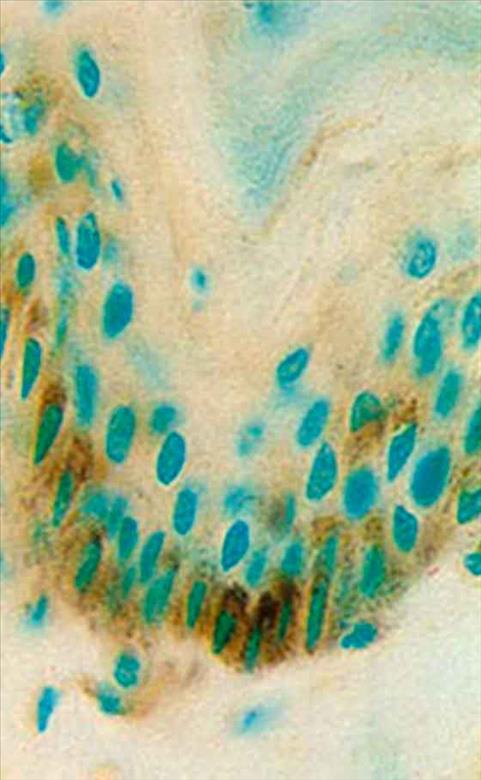
Human skin. 25 year old man
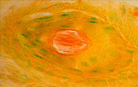
X 1250. Acid phosphatase reaction, 70 minutes. (© 1995 Vevy Europe Photoimage)
Section of hair follicle. The greatest reactivity is in the medulla. Nucleous stained in green
Skin of a guinea-pig
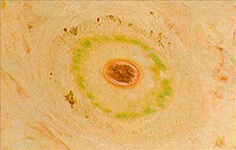
X 500. Acid phosphatase reaction, 50 minutes. (© 1995 Vevy Europe Photoimage)
Section of hair follicle. As in human subjects (Fig. 7-8) the activity is localized in the medulla that appears stained in red for the presence of pararosaniline and it is a lso stained in brown for the presence of melaninic pigment.
Skin of a guinea-pig
X 125. Acid phosphatase reaction, 50 minutes. (© 1995 Vevy Europe Photoimage)
In the centre of the photo we can see the hair stalk. The reaction is visible in the external root sheath. In this section there are many dermal papillae. The stratum germin ativum is marked by many melanocytes with their dark pigments. In the derma there are many macrophages stained in red by the reaction.
Human skin. 80 year old woman.
X 500. Acid phosphatase reaction, 50 minutes. (© 1995 Vevy Europe Photoimage)
Section of a hair follicle. Here the reaction is in the medulla of the hair marked by a red stain. The other layers of the hair are visible thank to methyl green wh ich marks the nucleous of cells.
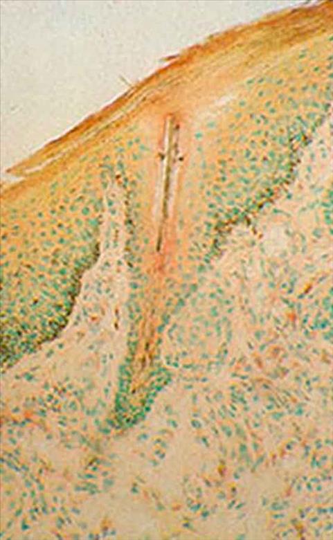
Human hair
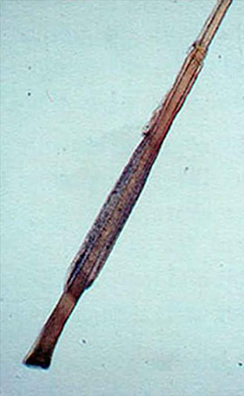
60 min. SD reaction. (© 1995 Vevy Europe Photoimage)
The pulled out hair has a sheath of epidermal cells adhering to the shaft and still fully active. The SD reaction, indicated by blue dots, proves this. With time, these cells die and gradually lose the enzyma tic activity investigated here. Activity decrease depends on time and it can be employed for biological dating. If applied when the hair is pulled out, this method shows hair functional condition and any related pathologies. Magn. 25x.
Cryostatic section of mouse miocardium.
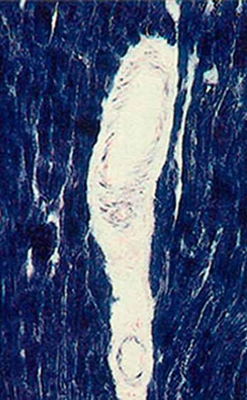
20 min. SD reaction. (© 1995 Vevy Europe Photoimage)
Cryostatic section of mouse skin.
60 min. SD reaction. (© 1995 Vevy Europe Photoimage)
In this low magnification picture the following elements can be identified from bottom to top: a layer of muscle tissue, in blue; just above it deep derma with a good number of longitudinally arranged (rose-shaped) fibroblasts; then the surface derma and pilosebaceous complexes with SD activity and finally the epidermis. In the skeleton muscles, at the bottom, several cell groups can be identified having different blue intensity. This is due to a non simultaneous contraction of muscle fibres which, therefore, have different energy requirements and enzymatic complement. Fibre contraction is limited in time. During the resting stage, adjacent fibres contract. Thus the strong blue staining characterizes a fibre under contraction, whereas lighter colours indicate non contracted fibres. Magn. 125x.
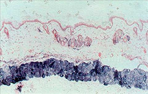
Cutis of woman aged 20
(© 1995 Vevy Europe Photoimage)
ATPase reaction after 5 minutes. Epidermis. The alignment of the germinative cells on the basement mambrane can be observed. The cells in the centre are stained blackissh-brown by the lead sulphide precipitate located in the ATPase activity site, in the apical zone of the cell. Magn. 1250x.
Skin of a 50 years old man
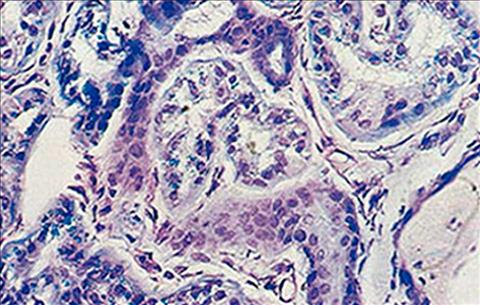
(© 1995 Vevy Europe Photoimage)
X 1250. Giemsa staining. Sweat eccrin gland. The section cuts many tubules. There are visible somewhere metacromatic and birifrangent granules in the epitheliocytes of the secretory part