Atlas
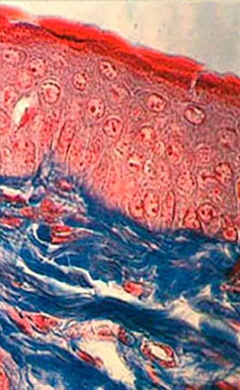
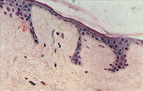
Cutis of a guinea pig
(© 1995 Vevy Europe Photoimage)
Staining Azan Mallory, Magn. 500x.
This methodology requires the utilization of two dyes: nuclear red and blue selectively staining the collagen fibers. The photo shows the uniformely pink stained epidermal layer and the bright red horny layer, whereas the dermis is blue stained. So me reddish fibroblast nuclei and endothelial cells pertaining to the capillaries of the dermis can also be observed.
In this picture, it is interesting to observe that the basal layer is entirely consisting of cylindrical cells, while the spinous layer has pcrfectly round nuclei, clearly featuring densifled chromatin nuclei (forming tiny red dots), most of which r epresent RNA messenger segments and, to a lesser extent, DNA segments.
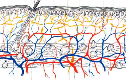
Distribution pattern of the arterial, venous and lymphatic plexus of the skin.
(© 1995 Vevy Europe Photoimage)
- Papillary capillaries;
- dead-end starting lymph capillaries;
- lymph branch of the superficial lymphatic plexus;
- candelabrum-shaped communicating branches debouching into the dermal papillae;
- super ficial venous plexus;
- hair with its follicle and its vascularized venous and artery branches of the superficial plexus;
- primary arteries and veins of the vasal plexus profundus;
- lymph collector of the lymphatic system of the superficial plexus;
- primary artery branch originating the artery plexus profundus;
- venous and artery branches descending towards the subcutaneous adipose lobuli.
In the papillary section, the lymphatic system is a closed systerm: consequently, the lymph circulates, in the first instance, outside the lymphatic system, directly bathing the dermic elements: this is the "plasmatic circulation" whi ch constitutes the internal means allowing nutritional exchange to take place. The plasmatic circulation which regulates the lymphatic circulation, is under the influence of the blood circulation; its exudation is regulated by blood pressure and by the os motic pressure of fluids, by nervous influences, endocrine influences, cellular metabolism, by the state of constriction and dilation of the vessels, and finally by the release of H vasodilatory substances emitted in large amounts by irritated cell tissue .
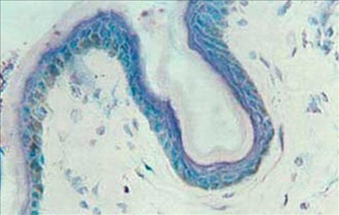
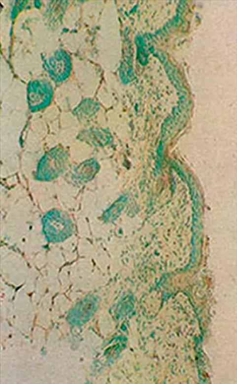
Cutis of woman aged 45
(© 1995 Vevy Europe Photoimage)
Cutaneous fold with rather thin epidermis. This stain not only shows the cellular structure but also melanin which turns green due to metachromasia typical for this stain. Almost all the epithelial cells of the basal layer in this skin segment feature this pigment. It is not limited to melanocytes but also extends to normal epithelial cells which have ahsorbed the pigment from the latter. The skin indeed is tanned.
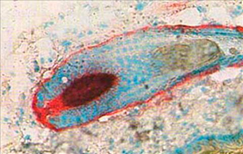
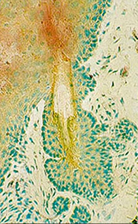
Human skin
(© 1995 Vevy Europe Photoimage)
Histochemical reaction to localize enzyme alkaline phosphatase activity. Reactivity is essentially localized in the dermal hair component (papilla and cuticle), since keratinocytes are deprived of such enzyme. The capillaries are red stained, especially those in active proliferation. The dermal papilla, rich in alkaline phosphatase, is sranding out because the hair section is in anagen phase in which this organule is in its highest development and trophism stage. Magn. 300x
Cutis of 80-year-old man
(© 1995 Vevy Europe Photoimage)
Magn. 500x.
Skin of a 50 years old man
X 500. Alpha-naphtyl reaction 30 minutes. (© 1995 Vevy Europe Photoimage)
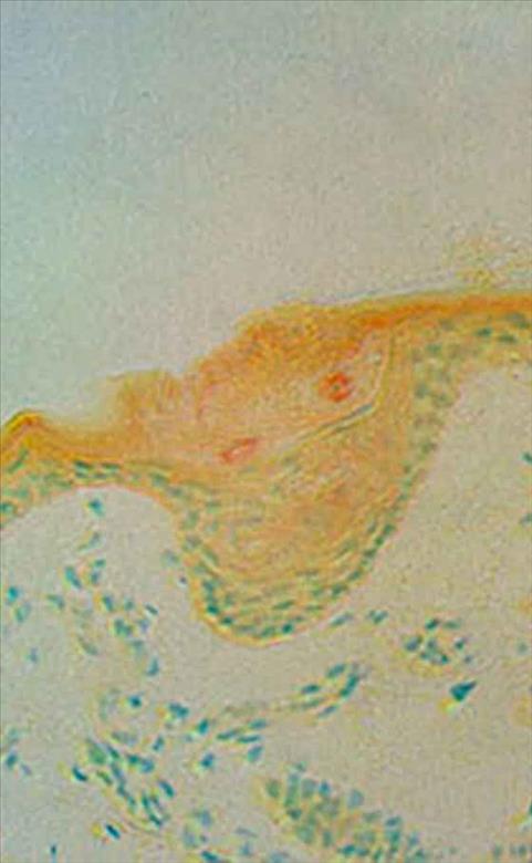
Cutis of a 70 days old mouse treated for one week with Oil-1
(© 1995 Vevy Europe Photoimage)
The almost complete absence of enzymic activity may be noticed as shown by the red (pararosaniline) dye, both in the horney layer and in numerous piliferous formations in the derma. You may notice the absence of keratin matter (dandruff) in exfoliation phase. Acid phosphatase reaction, 90 minutes, 200x.
Human skin. 80 year old woman
X 1250. Acid phosphatase reaction. (© 1995 Vevy Europe Photoimage)
Pararosaniline fixes in the place where the enzymatic activity is and stains with a red colour the deepest cells of the stratum corneum. Note the evident differenc e between weak stained epidermis, compared with the stratum corneum, and dermis completely unstained by the eaction.
Human skin. 80 year old woman
X 200. Acid phosphatase reaction, 70 minutes. (© 1995 Vevy Europe Photoimage)
Superficial part of sweat glands. You can notice an intense acid phosphatasic activity around the two pores (two little red rings). The positive elements belong to the stratum corneum
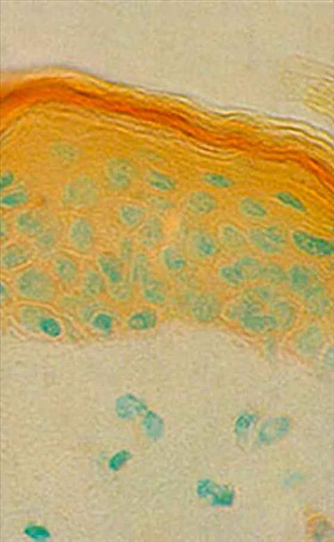
Untreated cutis of a 70 days old mouse
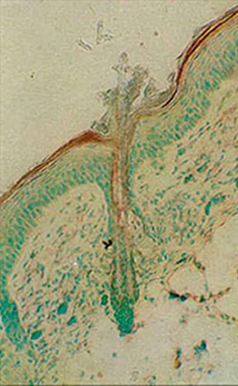
(© 1995 Vevy Europe Photoimage)
The brownish-red colour shows the localized reaction of the horny layer, the green coloured cell nuclea are revealed by a contrast dye. You may notice the two piliferous twin formations originating fro m the same dermal papilla. The reaction involves the two hair shafts. Acid phosphatase reaction, 80 minutes, 300x.