Atlas
Skin of a 50 years old man.
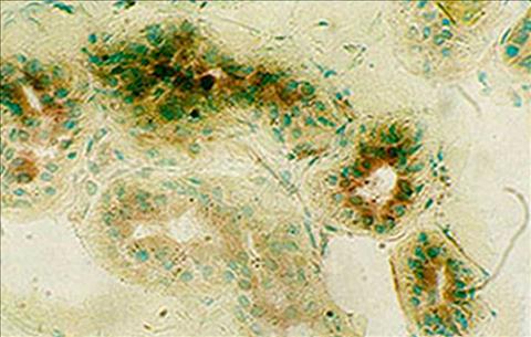
(© 1995 Vevy Europe Photoimage)
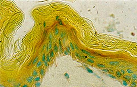
X 1250. Alpha-naphtyl esterase activity, 30 minutes. With a greatest magnification it is visible the difference between the activity of the secretory portion (down) and these of the escretory duct (up). The last c an be distinguished for its greatest activity evidentiated by a dark brown staining. Green stain of the nucleous is due to methil green.
Skin of a 50 years old man
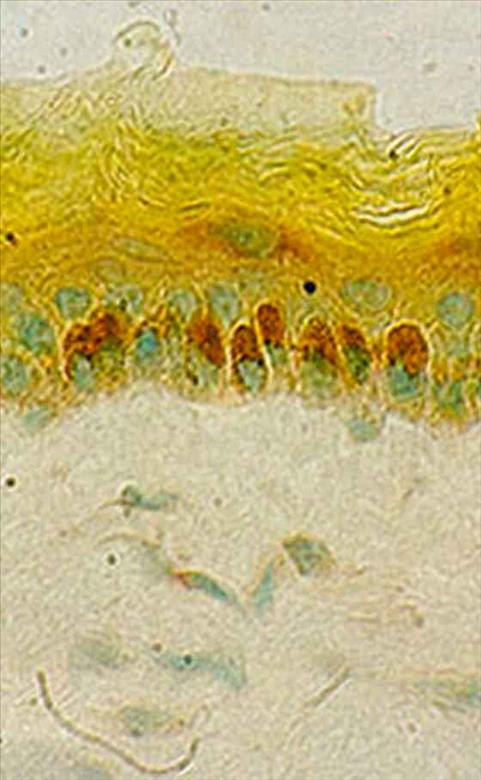
(© 1995 Vevy Europe Photoimage)
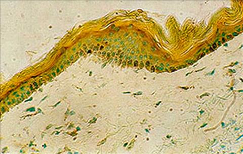
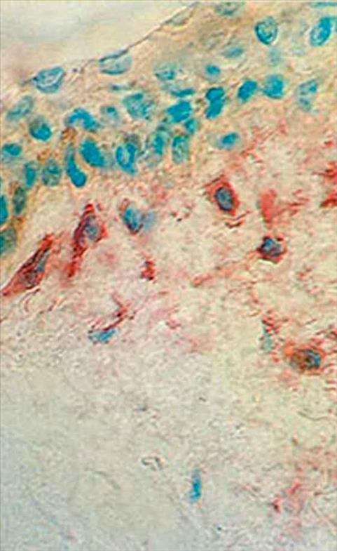
X 1250. Alpha-naphtyl esterase reaction, 20 minutes. Group of about ten melanocytes with melaninic granules. The stratum granulosum is stained with pararosaniline which evidentiates the places were the enzymatic a ctivity is
Mast cells or Mast-zellen.
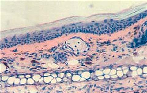
(© 1995 Vevy Europe Photoimage)
Giemsa Staining. Magn 300x.
Ear section from normal mouse (skin of Balb/C) A line of globoid cells with large empty spaces inside can be observed at the bottom. They are chondrocytes, i.e. cartilage cells of the pinna. At the top, the skin can be seen, stained in pink, with s ebaceous glands (in the middle), hairs, fibroblasts and mast cells. The latter cells are easily detectable because of the purple colour they acquire when stained with Giemsa, due to metachromasia. In this specimen, mast cells have a normal shape with a homogeneous distribution in the skin. They are in a patrolling state, though no inflammation is under way. The purple stain of these cells is expressed by va soactive substances contained in their vacuoles and released when stimulated by antigens or inflammatory factors. Rashes, burning and swelling of inflamed parts are mainly due to these cells that, with their substances, amplify the response against harmful agents. They allow greater blood flow to supply the damaged area with abundant plasma proteins to ensure immune defense.
Skin of a 50 years old man.
(© 1995 Vevy Europe Photoimage)
Skin of a 50 years old man
(© 1995 Vevy Europe Photoimage)
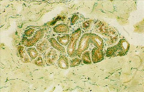
X 500. Alpha-naphtyl esterase activity, 30 minutes. Sweat eccrine gland of the clavear area. The ducts are composed of a double layer of cuboid cells marked by the activity. Above we can see some duct sections mor e reactive. The cells here have a reabsorbtion tuch and perhaps they need a greatest quantity of enzyme. The enzymatic activity is connected with physiological cellular activity.
Skin of a 50 years old man
(© 1995 Vevy Europe Photoimage)
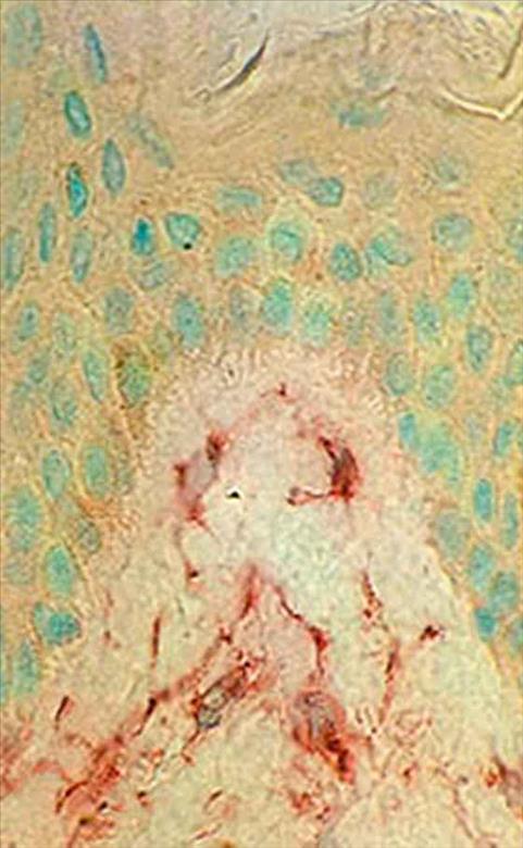
X200. Alpha-napthil esterase reaction 33 minutes. Epidermis and dermis. The enzimatic activity of skin is localized in the stratum granulosum which seems marked by a continuous red-brown strip. That means that th ese cells have a great production of enzyme, even if they are in their penultimate phase of life. We can also see many melanocytes in the stratum germinativum: here the staining is only due to their pigment. All nucleous are stained by methil green.
Skin of a 50 years old man
(© 1995 Vevy Europe Photoimage)
X 1250. Alpha-naphtyl reaction, 30 minutes. The section cuts an hair-sebaceous formation in eruptive phase. Activity is concentrated in the neck of the sebaceous gland and in the epithelial cells standing up the h air in growth phase, were the further hair duct will pass. Epiderm under hair push has bend. We can see down in the photo a sweat gland.
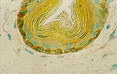
Skin of a 50 years old man

(© 1995 Vevy Europe Photoimage)
X 500. Alpha-naphtyl reaction 20 minutes. Root of a developed hair. There is no activity in the epithelial cells of the hair duct. The allonged positive cells (brown with green nucleous) along the hair are macroph ages. They are disposed in a strategical way in the point of less cutaneous defence; in fact hair determin an interruption of the dermic barrier. We can easily see some melanocytes in the hair bulb. Down in the photography is a sweat gland.
Cutis of a man aged 60
(© 1995 Vevy Europe Photoimage)
LAP reaction after 60 minutes. Dermal papilla. Some fibroblasts with strongly reacting long cytoplasmatic fibers are clearly visible. No LAP activity in the epidermis. Magn. 1250x.
Cutis of a woman aged 55
(© 1995 Vevy Europe Photoimage)
LAP reaction after 50 minutes. Epidermis and dermis: intense red granular (lysosomial) reaction of the dermal fibroblasts. This enzyme proves the existence of an intensive peptidase activity in these elements derivi ng from complex proteolysis and proteosynthesis phenomena. Magn. 1250x.